The Science Hidden in a Drop of Blood
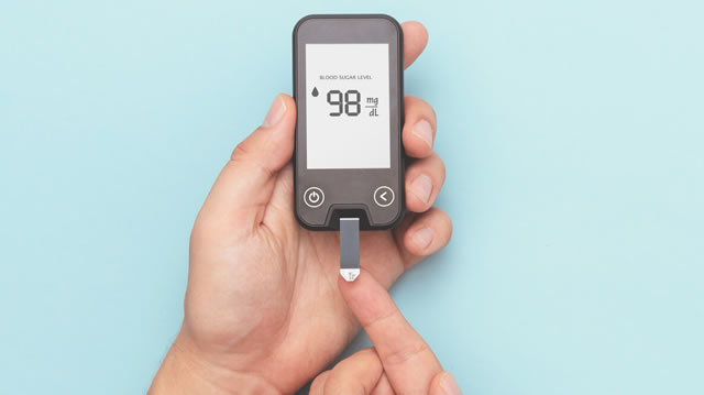
For decades, measuring blood glucose required full laboratory tests, blood collection tubes, and hours of waiting before results were available. Today, millions of people can check their glucose levels in just a few seconds using a small portable device and a single drop of blood taken from the fingertip.
What appears to be a simple gesture — placing a drop of blood on a strip and waiting a few seconds — actually involves a sophisticated combination of chemistry, physics, electronics, and microengineering. Inside a simple disposable test strip lies a technology capable of identifying specific molecules, generating microscopic electrical signals, and converting them into readable numbers.
This technology has allowed people with diabetes to monitor their blood sugar daily, preventing serious complications and dramatically improving quality of life. But how exactly can such a small device “know” how much sugar is present in the blood?
The answer involves highly specific chemical reactions, extremely sensitive electrochemical sensors, and microscopic engineering capable of manipulating almost invisible volumes of liquid.
The basic principle: measuring glucose through a chemical reaction
Glucose is a type of sugar present in the bloodstream that serves as the main source of energy for the body’s cells. When glucose levels rise or fall too much, the body can suffer serious consequences, especially in people with diabetes.
A blood glucose meter, commonly called a glucometer, works by transforming the presence of this molecule into a measurable electrical signal.
To make this possible, the system uses enzymes that are highly specific to glucose. These enzymes are biological molecules that act as catalysts, accelerating chemical reactions and interacting only with certain substances.
In modern glucose meters, the enzymes most commonly used are:
-
glucose oxidase
-
glucose dehydrogenase
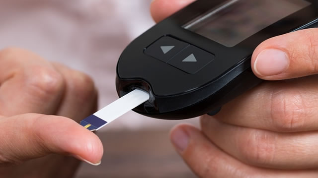
These enzymes are embedded in the test strip, which is the disposable component used for each measurement.
When the drop of blood comes into contact with the strip, the glucose present in the blood reacts with the enzyme. This reaction generates a transfer of electrons that can be detected by electronic sensors.
The fundamental principle is simple:
the more glucose present in the blood, the stronger the chemical reaction.
And the stronger the reaction, the greater the electrical signal produced.
The engineering hidden inside a test strip
At first glance, the strip used in a glucose meter looks like a small piece of plastic with metallic contacts. Internally, however, it contains a highly sophisticated structure.
Inside the strip there are several microscopic components arranged in layers:
-
microscopic channels through which the blood travels
-
a reaction chamber
-
enzymes specific to glucose
-
electron mediator substances
-
ultra-thin metallic electrodes
These microscopic channels are designed to guide a very small amount of blood to the area where the chemical reaction takes place.
This field of engineering is known as microfluidics, an area of science that studies how liquids behave inside microscopic structures.
Microfluidics is not used only in glucose strips. The same principle is also applied in pregnancy tests, rapid diagnostic tests, and devices known as lab-on-a-chip systems.
Why the drop of blood is “pulled” into the strip
One of the most noticeable things when someone uses a glucose meter for the first time is that the strip seems to automatically “suck in” the drop of blood.
In reality, there is no pump or suction mechanism inside the strip.
What occurs is a physical phenomenon known as capillary action.
Capillary action occurs when a liquid comes into contact with extremely narrow channels. Inside these channels two important forces come into play:
adhesion and cohesion.
Adhesion is the tendency of liquid molecules to stick to the walls of a material. Cohesion is the attraction between the liquid’s own molecules.
When the channel is extremely narrow, the molecules adhere to the walls and pull other molecules along with them. As a result, the liquid moves spontaneously through the channel.
The same phenomenon explains why:
-
a paper towel absorbs water
-
ink spreads through paper fibers
-
water can rise inside extremely thin tubes
In glucose strips, the microscopic channels are specifically designed to take advantage of this effect. As soon as the drop touches the opening of the strip, capillary action automatically pulls the blood into the internal structure.
Within milliseconds, the blood travels through the channel and reaches the reaction chamber.
The microscopic chamber where measurement happens
Inside the strip there is a tiny cavity called the reaction chamber.
This chamber has an extremely small volume and is designed to be completely filled by a very small amount of blood.
This detail is essential to ensure measurement accuracy.
If each test used different volumes of blood, the chemical reaction could vary and produce inconsistent results. For this reason, the strip’s design ensures that approximately the same sample volume is always used.
Inside this chamber are located:
-
the enzyme that reacts with glucose
-
mediator substances that transport electrons
-
electrodes responsible for detecting the electrical signal
As soon as the blood fills the chamber, the chemical reaction begins immediately.
The chemical reaction that reveals blood sugar levels
When glucose comes into contact with the enzyme present in the strip, an oxidation reaction occurs.
In simplified terms, this reaction causes the glucose molecule to lose electrons. These electrons are then transferred to mediator molecules present in the strip.
The mediator molecules carry the electrons to the metallic electrodes.
This flow of electrons creates an extremely small electrical current.
The strength of this current depends directly on the amount of glucose present in the sample.
If there is little glucose, few electrons are transferred. If there is a large amount of glucose, the electron transfer will be greater.
This principle converts glucose concentration into a measurable electrical signal.
Detecting nearly invisible electrical currents
The electrodes inside the strip act as electrochemical sensors.
They are capable of detecting extremely small electrical currents, often in the range of nanoamperes or microamperes.
To put this into perspective, these currents are millions of times smaller than the electrical currents used to power common electronic devices.
Even so, the glucose meter contains highly sensitive electronic circuits capable of measuring these tiny variations with precision.
The device then uses internal calibration algorithms to convert the electrical signal into a glucose concentration value.
This value is typically displayed in two units:
mg/dL (milligrams per deciliter)
mmol/L (millimoles per liter)
The entire process takes only a few seconds.
Why only a tiny drop of blood is needed
One of the major technological advances of modern glucose meters has been the dramatic reduction in the amount of blood required for testing.
Early devices required relatively large drops of blood. Modern meters, however, can work with volumes between 0.3 and 1 microliter.
For comparison, a typical drop of water may contain around 50 microliters. In other words, the amount used in a glucose test may be more than 100 times smaller.
Even so, this tiny sample contains an enormous number of glucose molecules.
Human blood contains billions of dissolved molecules in every microscopic fraction of liquid. Therefore, even a microscopic volume contains enough glucose to produce a detectable chemical reaction.
Thanks to the high sensitivity of electrochemical sensors, the device can convert this reaction into a reliable result.
The role of the microprocessor in producing the final result
Once the electrical current is detected by the electrodes, the signal is sent to the glucose meter’s electronic circuitry.
At this stage, the device’s microprocessor takes control of the process.
The microprocessor performs several steps in rapid succession:
measuring the electrical current intensity
correcting possible temperature variations
applying calibration curves
converting the signal into glucose concentration
These calibration curves are determined during the development of the device and ensure that the value displayed corresponds to the real glucose concentration in the blood.
Finally, the result appears on the device’s display.
Typical values may look like:
90 mg/dL
110 mg/dL
180 mg/dL
Depending on the clinical situation, these numbers may indicate normal, elevated, or reduced blood glucose levels.
Accuracy and limitations of glucose meters
Although they are highly reliable, home glucose meters are not as precise as laboratory tests performed in hospital equipment.
In general, home meters may present a margin of error of approximately 10 to 15 percent.
Several factors can influence the result, including:
ambient temperature
quality of the test strip
amount of blood applied
reaction timing
hydration status of the individual
Even so, these devices are considered sufficiently accurate for daily glucose monitoring.
In clinical practice, the trend of measurements over time is usually more important than a single isolated value.
Continuous glucose sensors: a new generation of technology
In recent years, even more advanced devices have emerged known as continuous glucose monitoring sensors.
These sensors are small devices applied to the skin, typically on the arm or abdomen. A microscopic filament sits just beneath the skin and continuously measures glucose present in the interstitial fluid.
Unlike traditional glucose meters, these sensors do not require a drop of blood for every measurement.
Instead, they monitor glucose levels continuously throughout the day and transmit the data to a reader or smartphone.
Although they rely on similar electrochemical principles, these systems represent a major advancement in the way glucose monitoring is performed.
A microscopic technology that transformed medicine
At first glance, a glucose meter may seem like a simple device. In reality, it combines complex scientific principles from several disciplines:
biochemistry
electrochemistry
microengineering
microfluidics
high-sensitivity electronics
Inside a small disposable strip there is essentially a microscopic laboratory capable of detecting specific molecules, generating electrical signals, and converting them into useful health information.
This technology has allowed millions of people to take an active role in managing their own health.
Instead of relying exclusively on occasional laboratory tests, people with diabetes can track their glucose levels daily and adjust diet, medication, and lifestyle with far greater precision.
What once required an entire laboratory can now fit in a pocket — and it works thanks to an elegant combination of science, engineering, and applied chemistry.
All of this happens in just a few seconds, starting from something almost invisible: a single drop of blood.